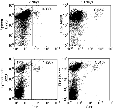
Figure 7.
CD40L-activated B cells efficiently home to secondary lymphoid organs. Single-cell suspensions from GFP transgenic mice were cultured on CD40L-transfected L cells in the presence of rIL-4 and cyclosporin as in the legend to Fig. 1. Seven days later cells were washed and 3 × 10⁷ cells injected i.v. into C57BL/6 mice (four mice/group). Mice were killed 7 or 10 days later. Spleens and inguinal lymph nodes were removed, stained with PE-labelled B220-specific or isotype-control antibody and analysed by flow cytometry. Quadrants were set using PE–isotype antibody-treated C57BL/6 spleen or lymph node cells. Data presented were gated on lymphocytes, as determined by forward and side scatter parameters, and are representative of results obtained in each of four mice killed 7 and 10 days after cell injection. The percentage of cells falling into the B220+/GFP− and B220+/GFP+ upper left and upper right quadrants respectively are indicated.

