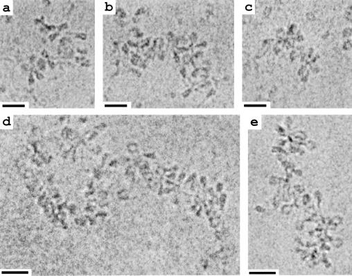
Figure 2.
EC-M of chromatin from COS-7 cells (a–c) vitrified in ≈40 mM ions and chicken erythrocyte nuclei (d and e) imaged in ≈15 mM ions. The fiber structure is consistent with an accordion-like compaction of the loose zigzags seen in Fig. 1. (Bar = 30 nm.)

