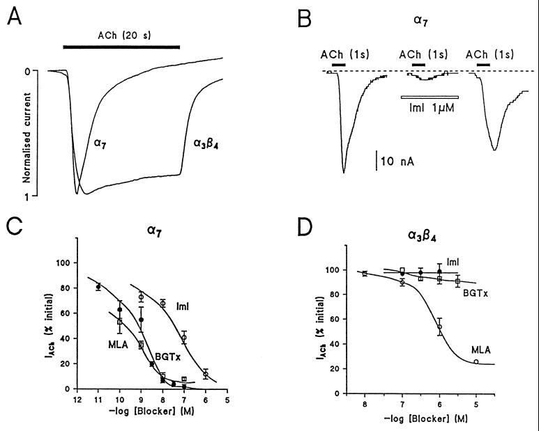
Figure 4.
Kinetics and blockade by α-toxins of IACh, in Xenopus oocytes injected with cRNAs for α7 or α3β4 nAChR. Oocytes maintained at a holding potential of −60 mV, were stimulated with pulses of ACh (100 μM) as shown by the top horizontal bars. (A) Two original IACh traces (normalized currents) obtained by stimulation with ACh (20 s) in one oocyte expressing α7 receptors and in another expressing α3β4 receptors. (B) Effect of α-conotoxin ImI on IACh generated by 1 s pulses of ACh (100 μM) applied to oocytes expressing α7 receptors. (C and D) Concentration-response curves of α-toxins to block IACh in oocytes expressing α7 nAChR (C) or α3β4 receptors (D). Protocols to estimate the blockade exerted by each toxin on IACh were as those described in B. Repeated pulses of ACh (1s, 100 μM) generated inward currents that were normalized to 100% (before adding each toxin concentration). The blockade exerted by each toxin concentration was expressed as percentage of the initial current. Data are means ± SE of 5–10 oocytes for each blocker.

