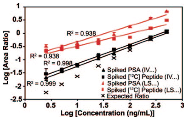
Fig. 7. Protein and 12C-synthetic peptide calibration curves for quantifying human PSA in nonfractionated, depleted plasma.

Calibration curves were generated with intact PSA added to depleted plasma prior to digestion and with 12C-synthetic peptides added to digested, depleted plasma. Two peptides, IVGGWECamcEK and LSE-PAELTDAVK, were used to quantify PSA by using the area ratio of light peptide to heavy peptide from the XICs of the 539.3/865.4 and 636.7/943.4 transitions, respectively. The concentration of 12C-peptide added was equivalent to the concentration of intact protein added. Three biological replicates for each concentration point were generated with intact PSA, whereas the calibration curves with 12C-synthetic peptides were generated once. Each point for all curves was analyzed in triplicate by MRM. Error bars indicate S.D. of the measurements.
