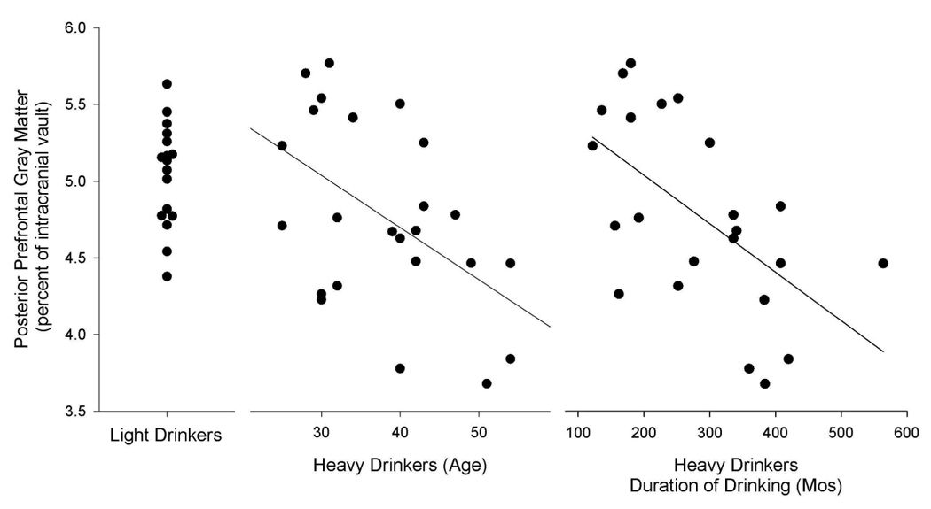
Figure 1.

posterior prefrontal gray matter (as a percent of the intracranial vault) for LD and HD. This is the regional variable with the largest difference between LD and HD. This figure illustrates both the volume reduction in HD versus LD, and the association in HD of this regional brain volume with both subject age and lifetime duration of alcohol use. We note that the data for HD is presented twice, once in relation to age and again in relation to duration of alcohol use.
