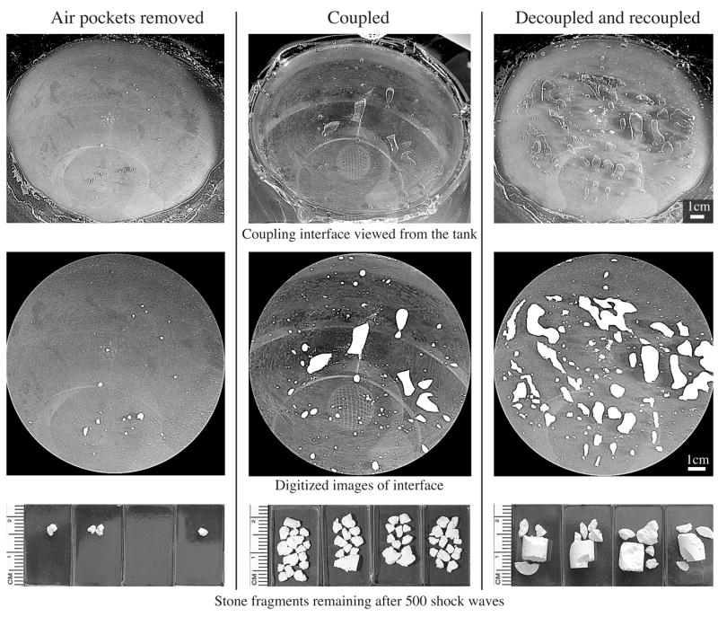
Fig. 3.
Three coupling regimes used for in vitro testing. When air pockets were manually removed from the coupling interface (left) stone breakage was more efficient than with single coupling (middle) or decoupling/re-coupling (right). Top row shows original photographs of the coupling interface. Middle row shows digitized images with air pockets highlighted for better visibility. Surface area occupied by air pockets in these images was about 0.5%, 6% and 15%, respectively. Bottom row shows stone fragments retained by 2-mm mesh basket following 500 SW’s (power level 3, 60 SW/min) for four stones treated with each coupling regime.

