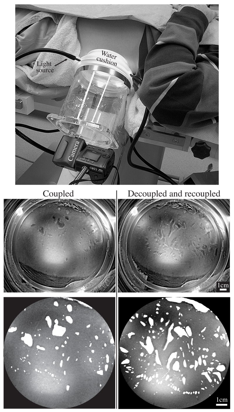
Fig. 5.

Air pockets became trapped when the lithotripter water cushion was coupled to skin. The cushion was filled with water and mounted on an acrylic cylinder so that the coupling interface could be viewed from behind. Similar to the in vitro setup (Fig. 3), air pockets became trapped against the skin with single coupling, and increased with decoupling/re-coupling (surface area occupied by air pockets in these representative images was about 8% and 16%, respectively).
