
Fig. 2.
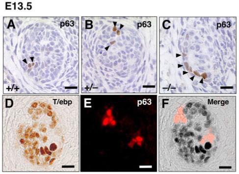
Expression of p63 and T/ebp in ultimobranchial body (UBB) cells. A-C: Transverse sections of UBBs from E13.5 wild-type, T/ebp-heterozygous and T/ebp-null mutant embryos. p63-positive cells are shown in brown. Dorsal is up. Only a few p63-positive cells can be detected per section in all genotypes, and a vast majority of UBB cells are negative for p63. D-F: Double staining for T/ebp (D, brown) and p63 (E, red) using wild-type UBB. Dorsal is up. F: The merged image demonstrates the complementary expression of T/ebp and p63 in the UBB. Scale bar = 20 μm.