Fig. 3.

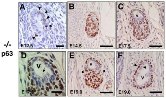
Altered ultimobranchial body (UBB) development and characteristic pattern of p63 expression in T/ebp-null mutants. A-F: Transverse sections of UBBs obtained from various embryonic days of T/ebp-null mutants were subjected to p63 immunostaining. Dorsal is up. Cells stained in brown indicate p63 expression. A: No significant difference was observed in the number of p63-positive cells between E12.5 and E13.5 UBBs (Fig. 2C). B: The p63-positive cells make a solid, oval mass by E14.5 that have taken over most of the UBBs. C,D: By E17.5, a vesicular structure is generated at the dorsal part of the UBB (v in C), which is lined by a monolayer of cells without p63 expression, accompanied by occasional ciliated cells (D, arrow). E: By E19.0, the UBB becomes constricted between the vesicular structure and the mass of p63-positive cells (shown by arrows). F: Cranial to the section shown in E, the vesicular structure displays a well-defined double layer of cells composed of p63-negative inner layer (arrowhead) and p63-positive surrounding layer (arrow). Scale bar = 20 μm in A,D, 50 μm in B,C,E,F.
