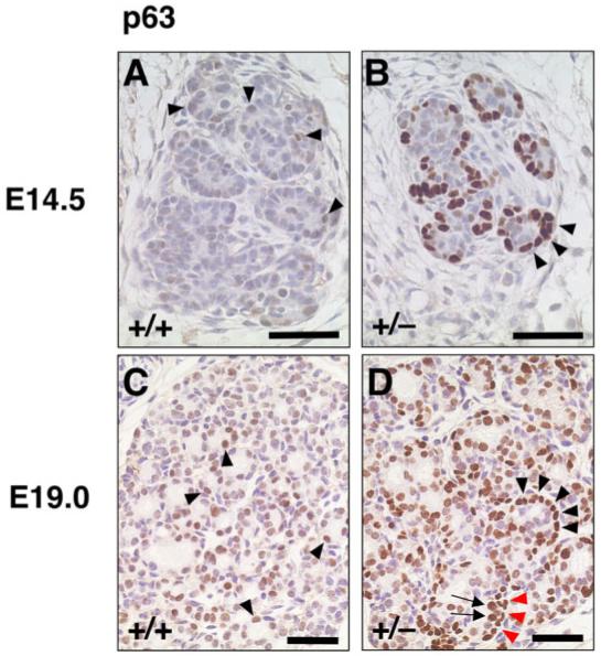
Fig. 5.

Immunostaining for p63 in primitive thyroid follicles of wild-type and T/ebp-heterozygous mutant embryos. A-D: The p63 expression is shown in brown. Dorsal is up. A,B: At embryonic day (E)14.5, the p63 expression level is mostly low or undetectable in primitive follicles of wild-type embryo thyroids (A, arrowheads), whereas almost all primitive follicles in T/ebp-heterozygous mutant thyroids are clearly rimmed with strongly p63-positive cells (B, arrowheads). C,D: At E19.0, many but not all follicular cells in primitive follicles of wild-type thyroid exhibit low p63 expression (C, arrowheads), whereas strong p63 expression is evident not only in follicular cells of primitive follicles (D, arrows) but also cells surrounding the primitive follicle (D, arrowheads) in T/ebp-heterozygous mutants, clearly showing a bilayered appearance in some part (D, arrows and red arrowheads). Scale bar = 50 μm.
