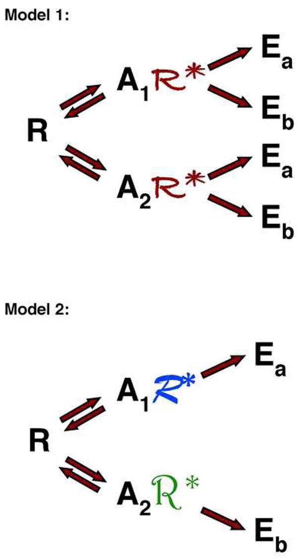
Figure 1. Models for receptor activation.

Model 1 shows two agonists (A1 and A2) that stabilize the same active receptor conformation (R*). The agonists differ in their binding affinity for the receptor and the effectiveness with which they can stabilize the active receptor conformation. However, since the conformation of the activated receptor (R*) is independent of the agonist bound, all agonists will stimulate the same spectrum of effectors (Ea and Eb) and produce the same spectrum of biological responses.
Model 2 shows two agonists (A1 and A2) that each stabilize a different active receptor conformation (denoted by R* in different fonts). Agonists differ both in their binding affinity for the receptor and the conformation of the activated receptor that they stabilize. Each receptor conformation will activate a characteristic subset of effectors (e.g. Ea or Eb) from the spectrum of possible effectors for the receptor and therefore each agonist can induce a characteristic set of biological responses that can differ from those induced by other agonists acting at the same receptor.
