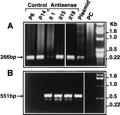
Figure 4.
PCR analysis of antisense and control transfected clones. PCR analysis of DNA from the selected clones was performed with primer pairs as described in Materials and Methods. (A) SP647 (sense primer) and the AP912 (antisense primer), both located in the CMV promoter (Fig. 3), were used to amplify a 266-bp fragment from cells transfected with the CMV promoter expression vector (antisense and empty vector transfected cells). (B) Sense primer SP647 (described above) and antisense primer SP7, located in the start codon region of PCDGF cDNA (Fig. 3), were used to amplify a 551-bp DNA fragment in the pAS-PCDGF transfectants only. No amplified band was obtained with DNA from control transfected cells. pAS-PCDGF plasmid DNA and genomic DNA from nontransfected PC cells were used as positive and negative control template for PCR, respectively.

