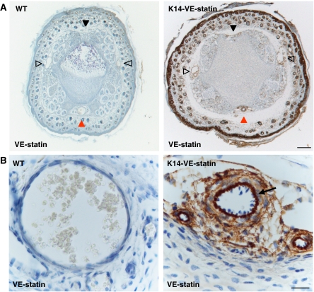
Figure 3.
Distribution of VE-statin/egfl7 in transgenic and WT animals. (A) Tail section of WT and transgenic P5 pups immunostained for VE-statin/egfl7. Solid black arrowhead: dorsal vascular bundle; open black arrowheads: lateral vascular bundles; red arrowhead: ventral vascular bundle (scale bar, 200 μm). (B) In the ventral vascular bundle, VE-statin/egfl7 accumulates between endothelial and smooth muscle cell layers (arrow) of the main artery (scale bar, 20 μm).

