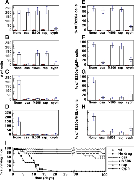
Figure 6. Suppression of Tumor Growth by Pharmacological Agents.
Tumor cells were harvested from lymph nodes and spleens and transplanted as described in Methods. The recipient mice were held until tumors became clinically apparent. Tumor recipient (open bars) and wild type (filled bars) mice then received daily injections of the indicated drugs for 7 d of either cyclosporine A (csa), FK506, rapamycin (rap), or cyclophosphamide (cyph). For (A–H), the mice were euthanized 24 h after the last injection of drug, and lymph nodes were harvested for analysis of either total number of cells (A–D) (expressed in single units representing 106 cells each) or surface markers of donor cells (E–H). For (I), the mice were observed over a span of 100 da and deaths recorded, as shown.
(A and E) Eμ-MYC tumors.
(B and F) Eμ-MYC/BCRHEL tumors.
(C and G) Eμ-MYC/BCRHEL/sHEL tumors.
(D and H) MMTV-rtTA/TRE-MYC/BCRHEL/sHEL tumors.
(I) Survival of animals bearing Eμ-MYC/BCRHEL/sHEL tumors. The statistical significance of the differences observed in the kinetics of mortality between the tumor-bearing mice that were either untreated, or treated transiently with cyclophosphamide is 0.01. The statistical significance of the difference in the mortality curves observed between those two groups and the tumor-bearing mice treated with either of the three immunosuppressant drugs is p < 0.001.

