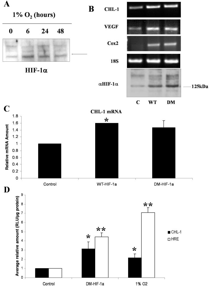
Figure 2.
HIF-1α drives expression of chordin-like 1 in retinal pericytes exposed to hypoxia. A: western blot analysis of nuclear extracts generated from human retinal pericytes exposed to increasing periods of hypoxia (0, 6, 24, and 48 h) for HIF-1α shows upregulation of the protein by 6 h. B: Transfection of human retinal pericytes maintained in normoxia with expression vectors for HIF-1 α, C is control/empty vector, WT is wild type vector, WT-HIF-1 α, DM is double mutant vector, DM-HIF-1 α, using the transfection reagent Fugene6, induces expression of many of the genes upregulated in response to hypoxia, as measured by RT–PCR. 18S PCR is shown as a loading control and western blot analysis confirmed expression from each of the HIF-1α expressing plasmids. C: Induction of CHL-1 mRNA in response to HIF-1α overexpression was quantitated by real time PCR. CHL-1 levels were normalized to 18S rRNA, using a pre-developed assay reagent. Data are expressed as mean relative quantity of mRNA, to control, for three independent experiments ±standard error of measurement. D: HeLa cells were transfected, using the transfection reagent Fugene6, with the CHL-1 promoter or a luciferase reporter construct containing four HIF-1α responsive elements (HRE), alone (control), cotransfected with the HIF-1α expression vector DM-HIF-1α, or alone and subsequent exposure to hypoxia for 24 h (1% O2). Cotransfection with DM-HIF-1α as well as exposure to hypoxia induced activation of the CHL-1 promoter and the HRE construct. Data are expressed as mean±SEM values. The asterisk indicates a significance at p<0.05 and the double asterisk indicates a significance at p<0.001.

