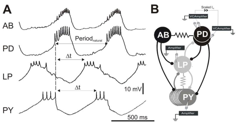
Figure 1. The pyloric network and experimental protocol.

A. Simultaneous intracellular recordings indicate that the AB and PD neurons oscillate in synchrony and inhibit the follower LP and PY neurons which burst out of phase with the AB/PD neurons and each other. The PD burst onset was used a reference point (dashed line) to calculate Periodnatural and the LP and PY burst phases. Phase is calculated by dividing the time difference between burst onsets ( t) by Periodnatural. The baselines for the membrane potential oscillations were −62 mV for AB, −57 mV for PD, −58 mV for LP and −62 mV for PY. For simplicity, recording of only one of two PD neurons is shown. B. The synaptic connectivity and the experimental paradigm represented schematically. All chemical synaptic connections (ball and stick symbols) are inhibitory; resistor symbols indicate gap-junctional couplings. All neurons were impaled with one electrode except for one PD neuron which was impaled with two for voltage clamp. The current used to voltage clamp that PD neuron was scaled and injected into the other PD neuron to voltage clamp the latter neuron to the same Vclamp value. Paired recordings of the LP and PY neurons were carried out in current clamp.
