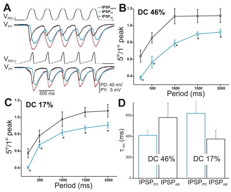
Figure 5. The IPSPs in the PY neuron in response to a train of two realistic PD waveforms.

A. The PY response to the stimulation of the PD neurons with DC 46% (top panel) and DC 17% (bottom panel) corresponding to amplitude 40 mV and cycle period 500 ms. B. Ratio 5th/1st peak in response to DC 46% plotted vs. cycle periods showed that IPSPAB depressed significantly less than IPSPPD (N = 6). Again, the amplitudes of the IPSPs were measured from Vrest. C. Similar to B, the extent of depression of IPSPAB in response to DC 17% was significantly less than IPSPPD. D. Average τrec values of the IPSPs calculated as in Figure 4E (N = 6). (* indicates significant difference).
