
Fig. 6.
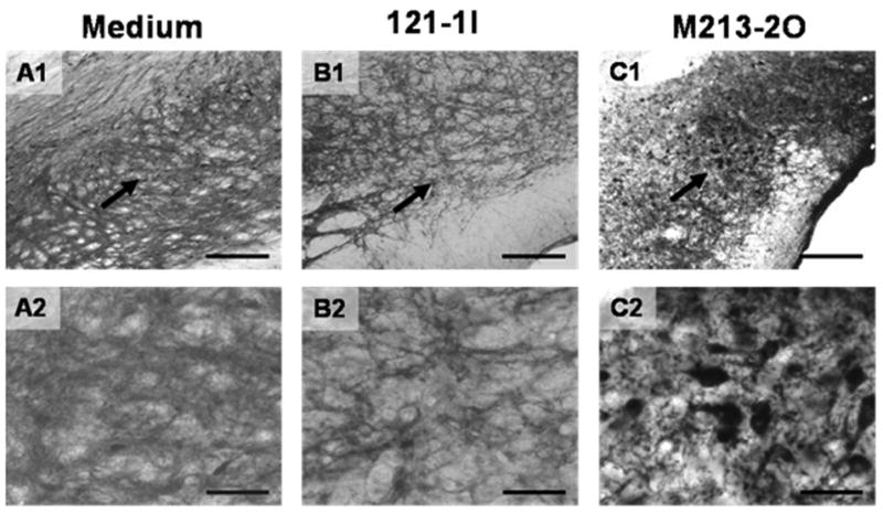
Immunohistological detection of glutamate decarboxylase protein (GAD) in coronal rat brain sections. Diaminobenzidine was used for chromogenic reaction. Shown are areas of the SNr either microinjected with medium (A1+2), or microimplanted with 121-1I cells (B1+2) or with M213-2O cells (C1+2). Arrows in the upper row (A1–C1) depict areas which are shown at higher magnification in the lower row (A2–C2). The tissues were treated identically and developed at the same time. Note the prominent positive staining for GAD in the GABAergic implants (M213-2O; C1+2), surrounded by immunopositive host cells, whereas in the other groups (A1+2; B1+2) only the positively stained host cells are visible. Scale bars 200 μm in A1–C1 and 50 μm in A2–C2.