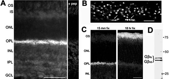
FIG. 1.
Gβ5 is present in the outer plexiform layer of the retina. (A) Immunofluorescent labeling of a mouse retinal section for Gβ5. All staining is blocked by pre-incubation of the antibody with the peptide immunogen (+ pep). (B) High-power view of Gβ5 labeling in the OPL. Labeling is associated with both rod and cone (arrows) terminals. (C) Fixation dependence of Gβ5 labeling demonstrated with retinas fixed for either 15 min or 18 h in 4% paraformaldehyde. (D) Western blot of mouse retina extract with the Gβ5 antiserum. Scale bars represent 50 μrn in A, 5 μrn in B, and 25 μrn in C. Abbreviations: OS, outer segments; IS, inner segments; ONL, outer nuclear layer; OPL, outer plexiform layer; INL, inner nuclear layer; IPL, inner plexiform layer; GCL, ganglion cell layer.

