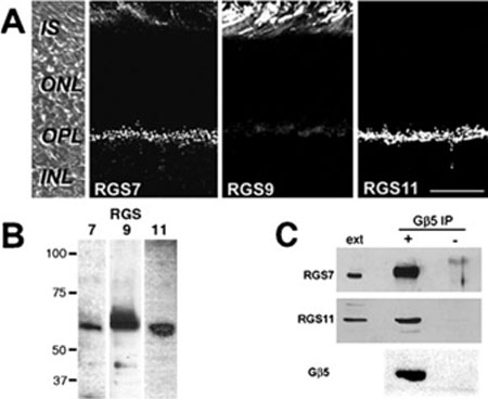
FIG. 4.
RGS7 and RGS11 are localized to the OPL and can be immunopre-cipitated from retinal extracts with Gβ5. (A) Mouse retina sections were labeled by immunofluorescence for RGS7, RGS9 and RGS11. The left panel shows a DIC image of a mouse retina section with the layers indicated. The scale bar represents 20 μm,and applies to all panels. (B) Immunoblots of mouse retinal extract with antisera against RGS7, 9 and 11. (C) Mouse retinal extract was immunoprecipitated with anti-Gβ5 (+) or preimmune serum (−), and then Western blotted for Gβ5, RGS7 and RGS11. Included on the blots were lanes containing retinal extract (ext). Abbreviations: OS, outer segments; IS, inner segments; ONL, outer nuclear layer; OPL, outer plexiform layer; INL, inner nuclear layer; IPL, inner plexiform layer.

