
Figure 2.
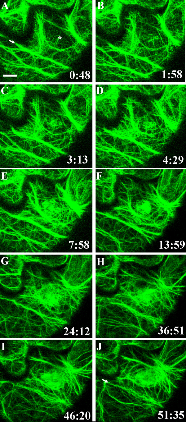
Formation of an actin patch beneath the contact site. Actin microfilaments visualized in the cortical cytoplasm underlying the outer epidermal cell wall in a cotyledon of A. thaliana expressing hTalin-GFP. The surface of the epidermal cell was touched with a glass microneedle at time 0:00 at the site indicated by the asterisk in A. Images A-J are projections of six optical sections taken at the times indicated in minutes and seconds. Actin microfilaments began to concentrate beneath the needle contact site 1 minute 58 seconds after touching the epidermal cell surface. The patch of actin continued to enlarge over the ensuing hour. The arrows in A and J indicate a thick actin cable that remains in the same position throughout the 51-minute sequence. A movie composed of images from 94 time points taken during this time is shown in Additional File 1. Bar = 10 μm.