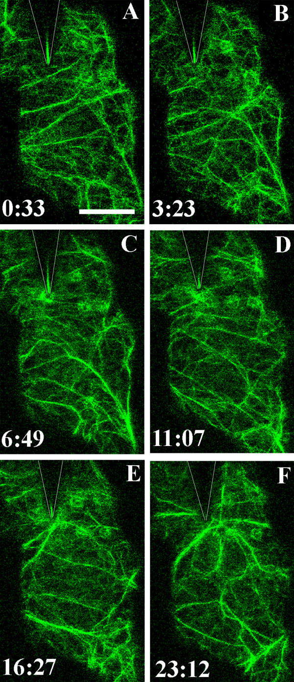
Figure 3.

Focusing of actin cables at the contact site. Actin microfilaments visualized in the cortical cytoplasm underlying the outer epidermal cell wall in a cotyledon of A. thaliana expressing hTalin-GFP. The surface of the epidermal cell was touched with a tungsten microneedle at time 0:00. The position of the needle is indicated by the shadow outlined by the V-shaped lines. The position of the tip of the needle is shown by a line of reflected light in many images. Images A-F are projections of six optical sections taken at the times indicated in minutes and seconds. Actin microfilaments began to concentrate beneath the needle contact site 3 minutes 23 seconds after touching the epidermal cell surface. The density of the actin patch fluctuated over the ensuing 20 minutes. Between 11–23 minutes after contact, actin cables became focused on the contact site. A movie showing focusing of the actin cables in this experiment is shown in Additional File 3. Bar = 10 μm
