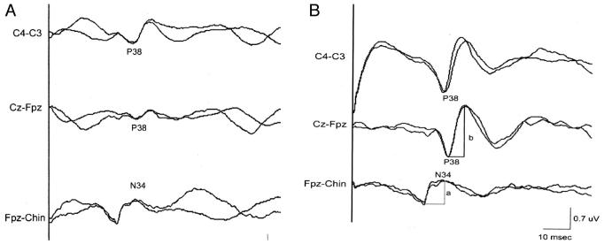
Figure 1.
A, right posterior tibial cortical SSEPs recorded under anesthetic conditions of treatment #1 (isoflurane 0.4% + N2O 70% + O2 30%). B, right posterior tibial cortical SSEPs recorded on the same patient under anesthetic conditions of treatment #4 (propofol 120 μg · kg-1 · min-1 + air + O2 30%). Measurements of N34 amplitude (a) and P38 amplitude (b) are illustrated.

