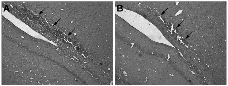
Figure 1.
Hematoxylin and eosin (H&E) stained paraffin sections of cortical contusions at 3 days post-TBI in vehicle and anti-CD11d treated animals. (A) Vehicle treated animal shows a large hemorrhagic contusion at the gray-white matter interface. (B) anti-CD11d treated animal has a much more focal area of injury after TBI. The upper boundaries of the contusion area are denoted with arrowheads (10X).

