
Figure 7.
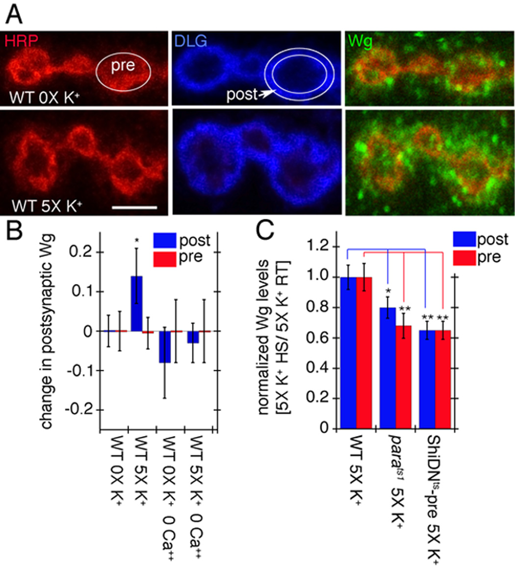
Activity-dependent Wg secretion by synaptic boutons. (A) Wg immunoreactivity (green) at synaptic boutons of wild type (top row) controls and (bottom row) specimens subjected to spaced 5X K+ depolarization in samples triple stained with anti-HRP (red), anti-DLG (blue) and anti-Wg (green). Images correspond to single confocal slices. The postsynaptic (DLG minus HRP) and the presynaptic (HRP) areas are outlined in white in the middle-upper and the left-upper rows respectively. (B) Pre- (red) and postsynaptic (blue) Wg levels in wild type controls and samples subjected to spaced 5X K+ depolarization, in the presence or absence of Ca++. Numbers in the Y-axis correspond to the difference in mean Wg intensity levels between control and experimental samples. (C) Pre- (red) and postsynaptic (blue) Wg levels in response to 5X K+ depolarization after blocking activity with parats1 and ShiDNts-pre. Wg levels were normalized by dividing the Wg levels after the 5X K+ paradigm at restrictive temperature (HS) by the Wg levels after the 5X K+ paradigm at permissive temperature (RT). Calibration scale is 2.5 µm in A.