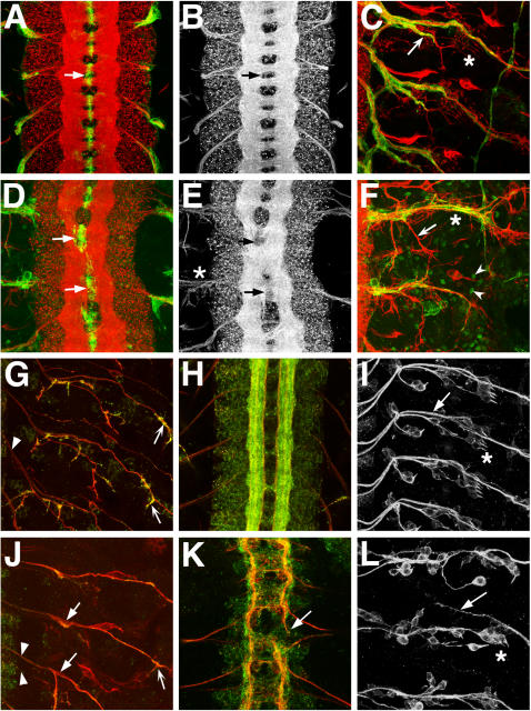
Figure 4. Phenotypes of wild type and Sec61α mutant embryonic nervous systems.
(A–C, G–I) Wild type. (D–F) Sec61αSH190 homozygous mutant. (J–L) Sec61αk04917 homozygous mutant. Anterior is to the top. PNS images have CNS off to the left in C,F,G,I,J,L. (A–C) Neurons are labeled with anti-HRP (red) and glia are labeled with mAb 5B12 (green). (G,H,J,K) Motor neurons labeled with mAb 1D4 (red) and anti-Synaptotagmin-1 (green). (I,L) Sensory neurons labeled with mAb 22C10. All embryos are late embryonic stage, approximately 22 hours old. (A) Wild-type CNS shows ladder-like pattern of axon tracts with commissural tracts ensheathed by midline glia (arrow). (B) Red channel (neurons) only of (A). (C) Wild-type PNS shows stereotypic pattern of motor- and sensory neuron projections (red) and coverage of nerve tracts by peripheral glia (green). SNa branch is indicated with arrow. (D) Sec61α embryos show disruption of CNS axon tract pattern (see also E), with glial profiles displaced compared to wild type (arrows). The confocal laser power relative to that used for imaging the wild type was increased to show glial staining pattern. (E) Neurons from (D) shown, highlighting lack of separation of central commissures (arrows). Defasciculation of peripheral axon projections at the CNS/PNS transition zone (asterisk) is also evident. (F) Sec61α mutant PNS shows variable disruption of axon patterning across hemisegments. Poor glial coverage of mistargeted SNa branch is indicated (arrow). Ectopic expression of mAb5B12 antigen is observed in the periphery (arrowheads). (G) Wild type PNS motor neuron pattern (red). Peripheral nerves near CNS/PNS transition zone are well fasciculated and tightly bundled (arrowhead). Synaptotagmin-1 is strongly detected at motor axon termini (arrows). (H) Wild type CNS motor neuron pattern (red) and distribution of synapse marker Synaptotagmin-1 (green). (I) Wild type PNS sensory neuron pattern. Arrow shows Anterior Fascicle sensory neuron tract pathway leading towards CNS on the left. (J) Sec61αk04917 mutant PNS motor neuron pattern (red) shows defasciculation (solid arrows) and lack of Synaptotagmin-1 immunolabeling (green) at motor axon termini (concave arrow) compared to wild type (G, concave arrows). (K) Sec61αk04917 mutant CNS motor neuron pattern (red) shows lack of development of CNS compared to same age wild type embryo (H). CNS axon pathfinding is disrupted along longitudinal connectives (arrow). (L) Sec61αk04917 mutant PNS sensory neuron pattern shows Anterior Fascicle sensory nerve aberrantly crossing hemisegment boundary anteriorly (arrow, compare to (I)). Round sensory neuron cell bodies are disorganized compared to stereotypic wild type pattern. (C, F, I, L) Asterisks indicate lateral chordotonal organs, which are disorganized in both Sec61αk04917 (L) and Sec61αSH190 (F) alleles compared to wild types (C, I).

