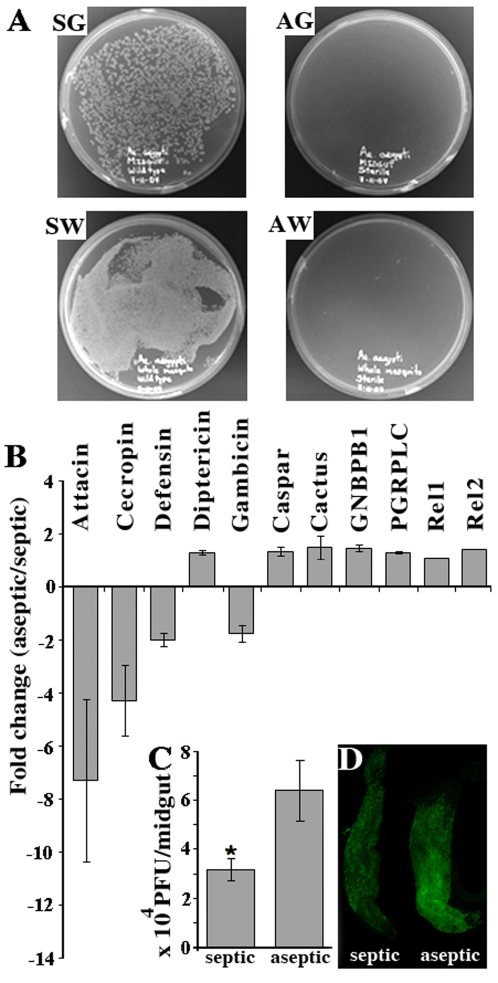
Figure 5. Elimination of the mosquito's endogenous bacteria reduces basal levels of immune gene expression and increases the susceptibility to dengue virus infection.
A. LB agar plates at 20 hours after plating homogenized and diluted septic gut (SG) and whole mosquito (SW) from non-treated mosquitoes, and aseptic gut (AG) and whole mosquito (AW) from antibiotic treated mosquitoes B. fold change in the expression of selected immune genes in aseptic mosquitoes compared to septic mosquitoes; C. virus infection levels in aseptic and septic mosquitoes were measured and compared by plaque assay in C6/36 cells * P<0.05 in Student's t-test. D. Dengue virus distribution and loads in septic and aseptic mosquito midguts assayed through IFA. Error bar represents standard error. Primary data for the real-time qPCR assays are presented in Table S2.

