
Figure 10.
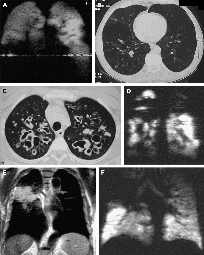
Examples of hyperpolarized 3-Helium MRI and correlation with HRCT. A, B Patient with alpha-1-antitrypsin deficiency. Notice basal ventilation defects on coronal MRI (a), with corresponding panlobular emphysema on axial CT (b). C, D Patient with cystic fibrosis. Notice upper lobe cystic bronchiectasis on axial HRCT (C) with corresponding ventilation defects on coronal hyperpolarized 3-He MRI (D). E,F Patient with lung cancer. On coronal proton image a large soft tissue mass is visualized in the right upper lung(E), which corresponds to upper lobe ventilation defect on hyperpolarized 3-He MRI (F).