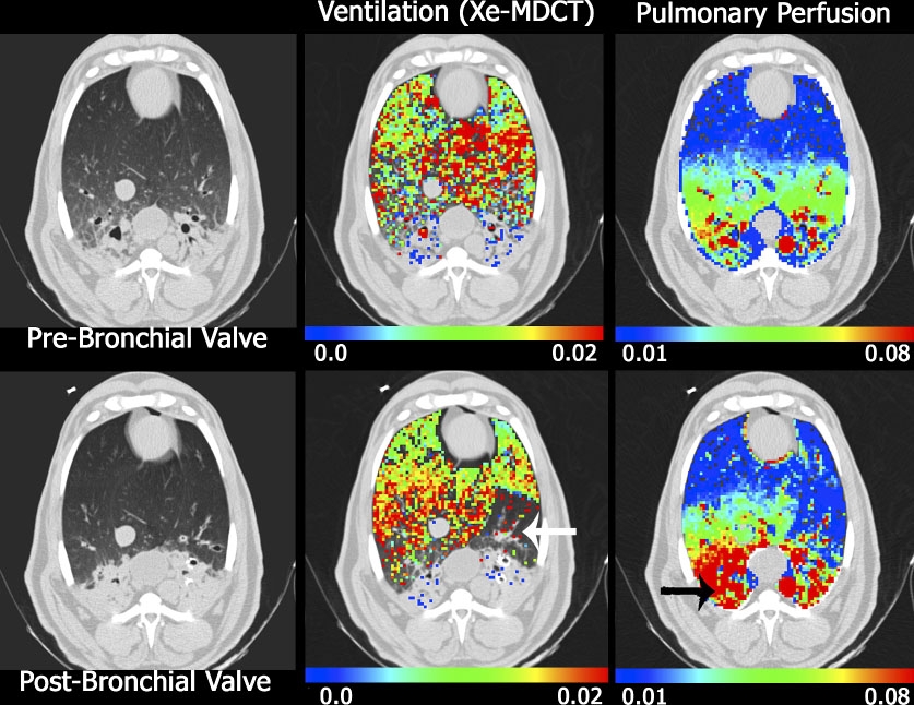
Figure 6.

Axial images from a sheep with native pneumonia (dependent lung regions), imaged supine, anesthetized in the MDCT scanner. Ventilation (middle column) and perfusion (left column) data sets were obtained before (upper rows) and after (lower rows) the placement of an endobronchial valve. The white arrow in the lower middle column marks the location of ventilation defect caused by the endobronchial valve. In this same region on the perfusion images (see lower right) there is a regional reduction in perfusion indicating regional, intact hypoxic pulmonary vasoconstriction. The black arrow in the lower right image marks a region that preferentially receives an increase in blood flow following the shunting of perfusion from the regional of the endobronchial valve, presumable because regional HPV is blocked in the presence of inflammation. Reprinted with permission from [74]
