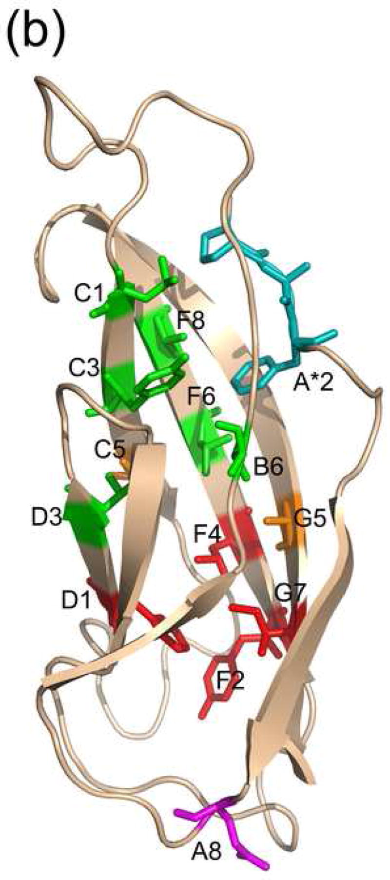
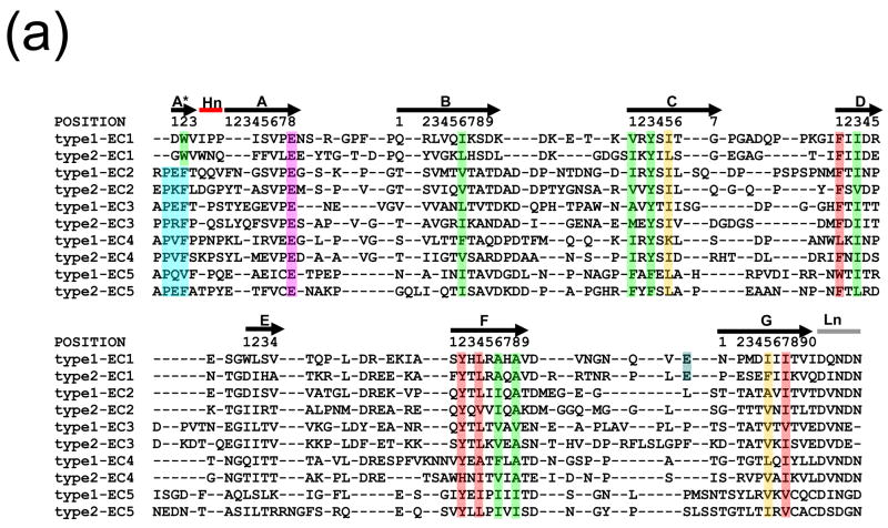
Figure 2.

(a) Structure-based sequence alignment of cadherin consensus sequences. Positions that are conserved in terms of residue type are highlighted, with residues of cluster 1 in green and residues of cluster 2 in red. The two bridge positions are in orange. Residue positions are labeled according to their positions in this alignment, using the typeI-EC1 consensus sequence as a reference. The EC1-specific position E89 is shown in blue, and the PxF motif of ECs 2-5 is in aqua. The hinge is denoted ‘Hn’ and the linker ‘Ln’. (b) The locations in the structure of the conserved positions in C-cadherin EC2 (PDB code 1L3W) are shown in stick representation and colored as in (a).

