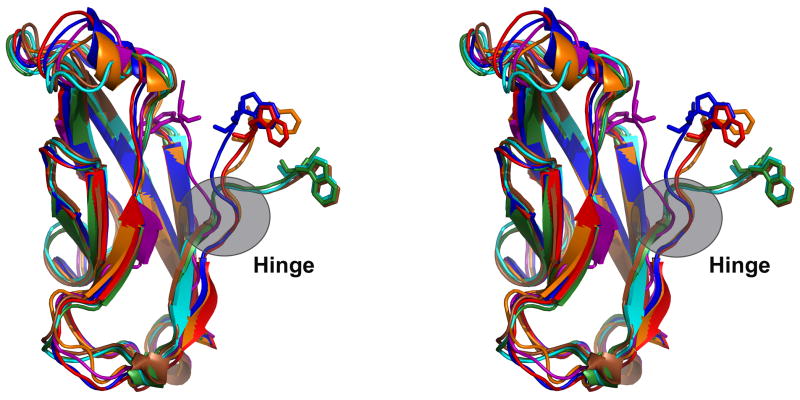
Figure 4.
Stereo view of alternate conformations of the A* strand. The E-cadherin EC1 monomer (PDB 1O6S) with W2 inserted into its pocket (purple) was aligned using Ska43 to a set of EC1 dimer protomers with W2 extended toward its partner molecule: E-cadherin (2O72, blue), N-cadherin (1NCG, orange), C-cadherin (1L3W, red), cadherin-8 (1ZXK, green), cadherin-11 (2A4C, cyan), and mn-cadherin (1ZVN, brown). The hinge connecting the swapping A* strand to the rest of the domain is indicated.

