
Fig. 1.
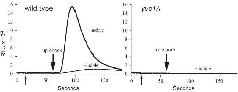
Indole potentiates up-shock-induced Ca2+ release through TRPY1 (Yvc1) in vivo. The luminescence of wild-type (left) or yvc1Δ (right) yeast cells expressing aequorin were examined. At the first arrow, nine volumes of either 0.5% DMSO (- indole curve) or 1 mM indole in 0.5% DMSO were added to the cells. Sorbitol was added at the second arrow at a final concentration of 1 M. Indole’s ability to potentiate the wild-type luminometric signal (in relative luminescence units, RLU) is evident (left). No response was seen in yvc1Δ cells (right) with the same indole treatment, indicating that the Ca2+ release and the indole effect are entirely due to the TRPY1 channel.