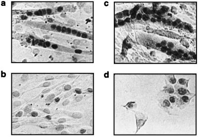
Figure 5.
Nuclear localization of Six1 and Six4 proteins. C2C12 cells (a and c, differentiated myotubes; b and d, proliferating myoblasts) were fixed on gelatin-coated slides with 4% formaldehyde at room temperature. Six1 and Six4 proteins were detected by 1:100 dilution of purified antiserum against Six1 (a and b) or 1:2,000 dilution of antiserum against Six4 (c and d). Horseradish peroxidase-conjugated goat anti-rabbit IgG (Jackson ImmunoResearch) was applied at 1:200 dilution, and then the immunoperoxidase reaction was used to reveal cellular localization of Six proteins. No signal was observed with the corresponding preimmune sera (not shown).

