
FIGURE 5.
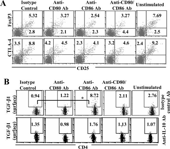
TGF-β1+ T cells, but not CD25+ or Foxp3+ T cells, are induced upon dominant co-stimulation by CD80. Co-stimulation blockade cultures were carried out as described in Fig 1. On day 5, cells from these primary cultures were washed, incubated with fresh medium for additional 48 h. A) These cells were stained for surface CD4 and CD25, and intracellular Foxp3 and CTLA-4, and analyzed by FACS. B) Purified CD4+ T cells from OVA primed mice were cultured as described above in the presence of additional antibodies (isotype control or neutralizing Ab against IL-10). Cells from these cultures were washed on day 5, cultured for an additional 48 h, and tested for CD4+ T cells positive for surface TGF-β1 by FACS. Samples were gated for CD4+ population for the panels A and B. Frequencies of CD4+CD25+Foxp3+ and CD4+CD25+CTLA-4+ cells are shown in panel A and CD4+TGF-β+ cells in panel B. These assays were repeated 4 times in triplicate with similar results and representative values are shown. * indicates a p value of <0.05.