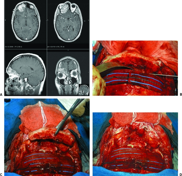
Figure 11.
(A) Magnetic resonance images of the brain show fibrous dysplasia of the orbital bar. (B) Operative photographs show the thickened orbital bar, which has been (C) osteotomized without violation of the medial canthal ligaments (level I transbasal approach). (D) The intraorbital exposure after removal of the orbital bar.

