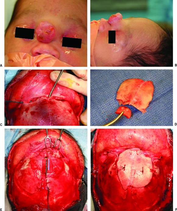
Figure 12.
(A) Anterior and (B) lateral photographs of a baby with a frontal encephalocele. (C) The lesion is outlined with a dissector after a bicoronal scalp incision is performed. (D) The frontal craniotomy flap. A split calvarium graft is used to reconstruct the nasion (level I transbasal approach). (E) After the encephalocele is resected, the bony defect involving the nasion is visible. (F) The defect is reconstructed with the bony fragments seen in (D).

