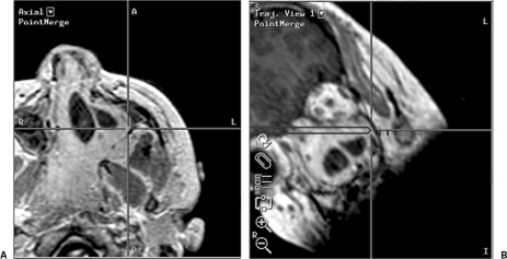
Figure 17.
(A) Axial localizing and (B) trajectory neuronavigational views show the microscopic point of focus to be located on the lateral wall of the maxillary sinus. This microscopic view was achieved during craniofacial surgery for resection of a large juvenile angiofibroma via a level III transbasal approach.

