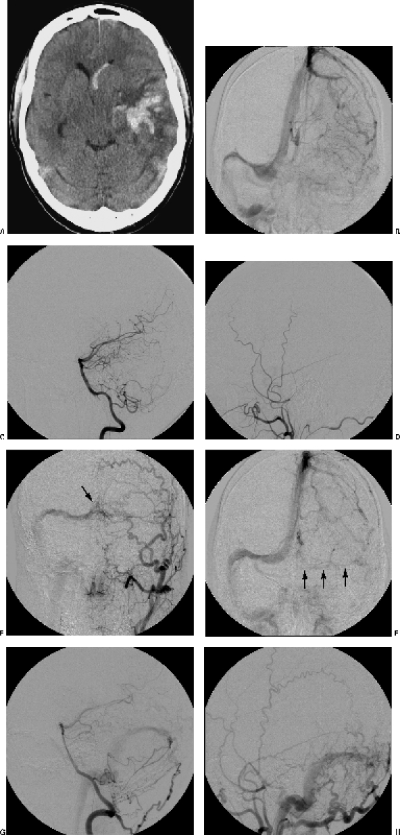
Figure 1.

(A) Preoperative computed tomographic image shows the left intracerebral and intraventricular hemorrhage. (B) Preoperative angiogram shows thrombosis of the left sigmoid sinus. However, the lateral projections of (C) the posterior circulation and (D) the left external carotid angiogram show no DAVF. Control angiograms 1 year after treatment show (F) partial patency of the left sigmoid sinus and (E,G,H) the formation of a DAVF between tentorial branches of the (E) left external carotid artery, (G) right vertebral artery, and (H) meningeal branches of the external carotid artery to (E) the right transverse sinus and confluens (arrow).
