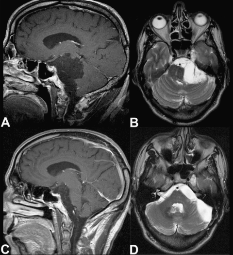
Figure 1.
Preoperative (A) sagittal T1-weighted and (B) axial T2-weighted magnetic resonance (MR) images show a CPA epidermoid mass dislocating the brainstem and extending to the prepontine region and temporal fossa. Postoperative (C) sagittal T1-weighted and (D) axial T2-weighted MRIs show the decompression of neurovascular structures and confirm removal of the infratentorial component. The lesion site has filled with cerebrospinal fluid. The right temporal remnant of the tumor was stable at the patient's 24-month follow-up examination.

