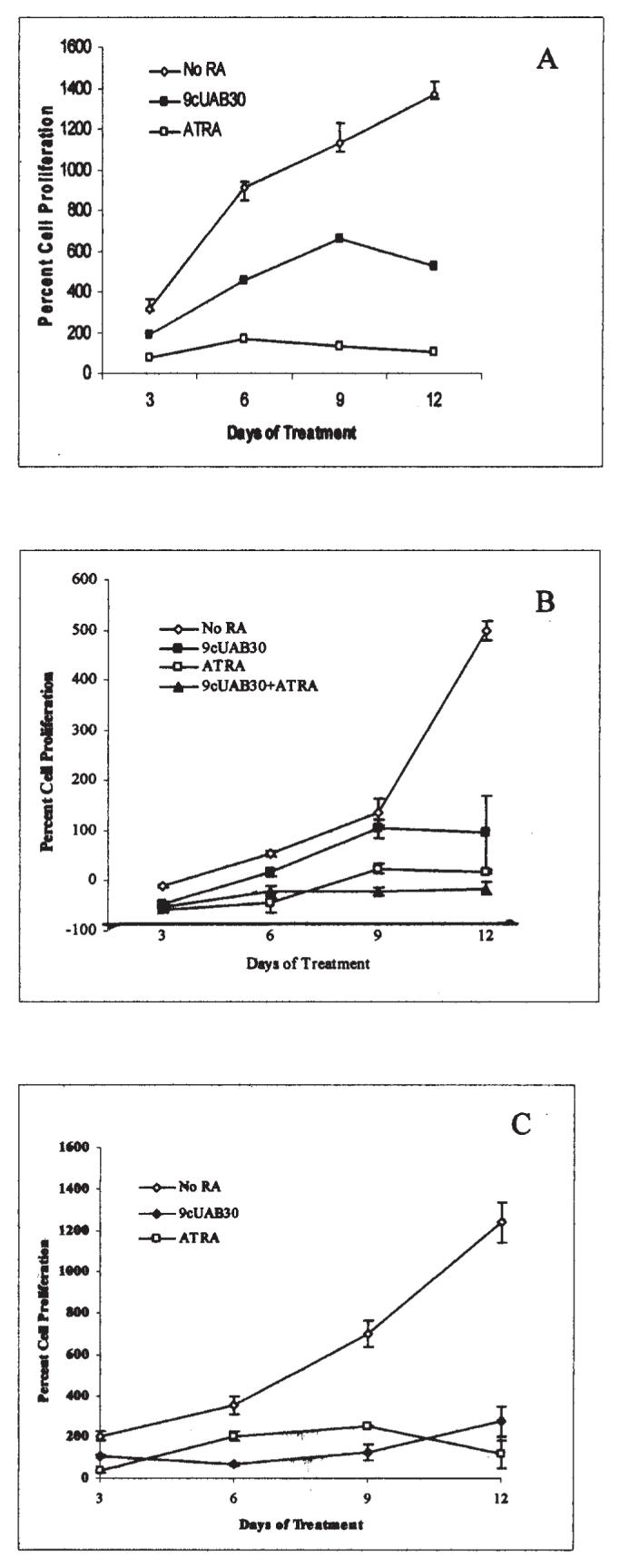
Figure 1.

Novel retinoid treatment decreases the proliferation rates of human breast cancer cells. (A) Human T47D breast carcinoma cells were grown in untreated control media (◇) or either 2 μM ATRA (□) or 10 μM 9cUAB30 (◆) for 12 days of concurrent culture. (B) Human MDA-MB-361 cells remained untreated (◇) or were treated with 10 μM 9cUAB30 (■), 2 μM ATRA (□) or a combination of 5 μM UAB-30 and 1 μM ATRA (▲). (C) Human MCF-7 cells were untreated (◇), treated with 10 μM 9cUAB-30 (◆), or treated with 2 μM ATRA (□). Proliferation is expressed as the percentage of the ratio of live cells harvested at days 3, 6, 9, and 12 of treatment to the number of live cells originally plated. Average cell numbers obtained by trypan blue exclusionary staining performed with a standard hemacytometer are presented with SEMs from triplicate culturing of each treatment group.
