
Fig. 1.
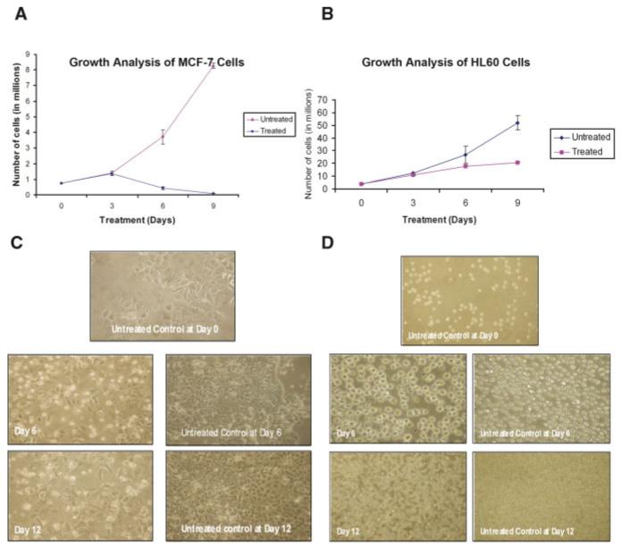
Growth analysis and morphological studies of MCF-7 breast cancer cells and HL60 promylocytic leukemia cells in response to EGCG treatment. Based on dose response analyses (data not shown), MCF-7 cells (A) were grown either in the presence or absence of 100 μM EGCG and HL60 cells (B) were grown in the presence or absence of 50 μM EGCG. Cells were counted using trypan blue staining. Morphological data were recorded at 3-day intervals for both MCF-7 (C) and HL60 cells (D). Cells were visualized at 100× magnification. [Color figure can be viewed in the online issue, which is available at www.interscience.wiley.com.]