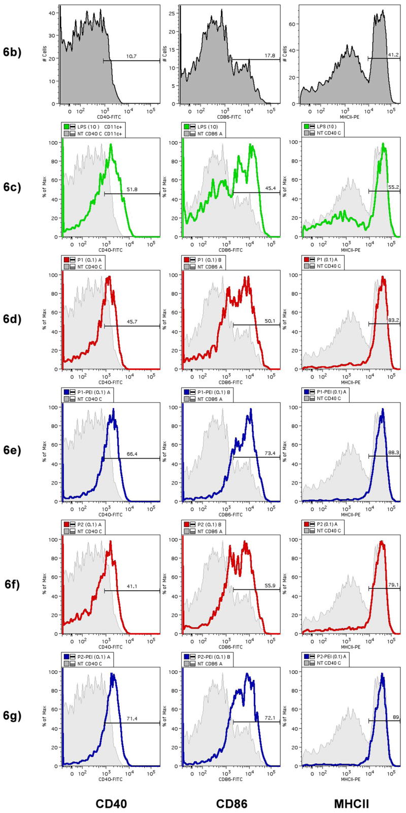
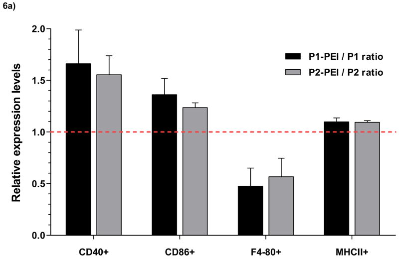
Figure 6.

POE microspheres containing plasmid DNA were incubated with primary CD11c+ B6 mouse bone marrow-derived dendritic cells (BMDCs). After 24 hours, BMDCs were stained for expression of activation markers CD40, CD86, and F4/80, which is down-regulated upon activation, and MHCII. Activation marker expression was analyzed by FACS. (a) Relative levels of expression of maturation and co-stimulatory markers after incubation with PEI-containing microspheres compared to incubation with non-PEI microspheres (n=3) expressed as a ratio of POE-PEI blended activation to unblended POE control activation. Values greater than 1 indicate increased expression of the surface marker with PEI blendding; less than 1 indicates decreased expression levels. (b–g) Histogram plots of expression of CD40, CD86, and MHCII expression after incubation with 0.1 mg/mL of POE microsphere. Untreated controls are shown in grey (b) and as background for comparison to incubation with LPS at 10 μg/mL (c), P1 (d), P1-PEI (e), P2 (f), and P2-PEI (g).

