
Figure 3.
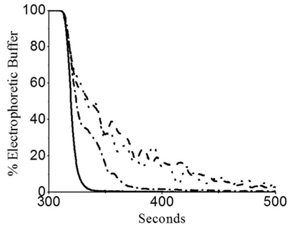
Decay of electrophoretic buffer flow upon closure of the elecrophoretic-buffer valve. For varying Δcapillary, the percentage electrophoretic buffer entering the capillary was measured over time. The tested Δcapillary were 0.75 mm (solid), 0.5 mm (dotted-dashed), 0.3 mm (dotted), and 0.1 mm (dashed). The physiologic buffer velocity: was 1.7 mm/s and the electrophoretic buffer flow rate was 0.1 ml/min.