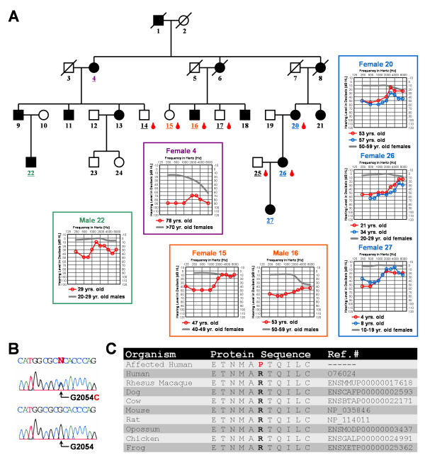
Figure 1.
Audiologic and genetic characterization of the low frequency hearing loss pedigree. (A) Each individual in the pedigree is assigned a number. Underlined numbers indicate that auditory evaluations were performed for that person. Affected individuals are denoted by blackened symbols, males are denoted by squares, females are denoted by circles, and deceased persons are indicated by a diagonal line through the symbol. Blood drop symbols indicate individuals who donated a blood sample to the study. Symmetrical hearing loss was detected in all affected family members; therefore, only the right ear pure-tone air conduction thresholds are plotted on the audiograms. Frequency in hertz (Hz) is plotted on the x-axis and the hearing level in decibels (dB HL) on the y-axis. Plotted on each audiogram (gray line) are the average pure-tone air conduction thresholds for a person with normal hearing matched in age [39] to the family member. (B) Electropherograms showing a heterozygous c.2054G>C mutated genomic nucleotide sequence from an affected individual compared to a homozygous c.2054G unaffected family member. Nucleotide numbering starts with the ORF. (C) Protein alignment shows conservation of the p.R685 residue during evolution. The p.685P substitution in the pedigree is shown in red.

