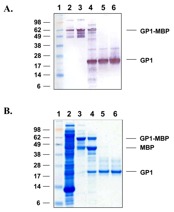
Figure 2.
Expression and purification of LASV GP1 from E. coli Rosetta gami 2 cells transformed with construct pMAL-c2x:GP1. An E. coli lysate was generated from IPTG-induced cells, the clarified supernatant was applied to an amylose resin column, the protein was eluted with 10 mM maltose, cleaved with Factor Xa, and purified by SEC. (A) Western blot of protein in (lane 2) whole bacterial cell lysate, (lane 3) amylose capture eluate, (lane 4) Factor Xa cleavage reaction, (lanes 5 and 6) SEC-purified GP1 generated from pooled GP1-containing fractions. The blot was probed with LASV mAb mix containing GP1-specific mAbs, then detected with an HRP-conjugated goat α-mouse IgG antibody. (Lane 1) Western blot XP standard molecular weight markers, with sizes (kDa) shown to the left of the panel. (B) SDS-PAGE and Coomassie blue stain of proteins in (lane 2) whole bacterial cell lysate, (lane 3) amylose capture eluate, (lane 4) Factor Xa cleavage reaction, and (lanes 5 and 6) purified GP1 generated from two sequential SEC runs. (Lane 1) SeeBlue® Plus2 pre-stained molecular weight markers, with sizes (kDa) shown to the left of the panel. GP1, MBP, and GP1-MBP are indicated.

