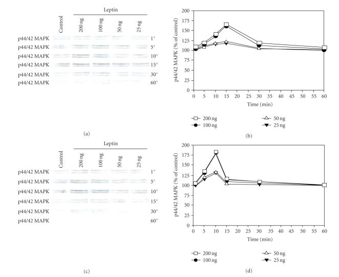
Figure 3.
Leptin stimulates MAPK phosphorylation in PC-3 and DU-145 cells. Growth factor-starved PC-3 (a) or DU-145 cells (c) were exposed to increasing concentration of leptin for various time points. Cell lysates obtained after each time point were separated on a 7.5% SDS-PAGE and Western-blotted. Phosphorylated MAPK levels were obtained after incubation with p44/42 MAPK antibody and chemiluminescent detection. (b) and (d), levels of p44/42 MAPK obtained after densitometric analysis and represented as percentage of control against time (control being set at 100%). The results of one of the three independent experiments are shown.

