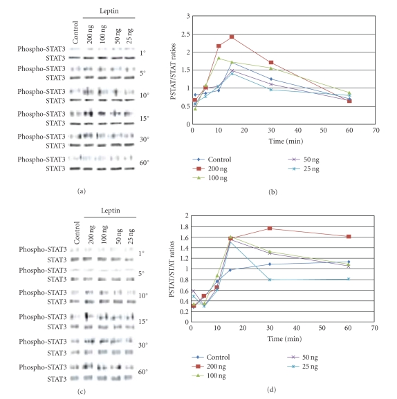
Figure 4.
Leptin stimulates early phosphorylation of STAT-3 in PC-3 cells. PC-3cells (a) or LNCaP cells (c), devoid of growth factors by an overnight exposure to basal medium, were exposed to increasing concentrations of leptin for various time points. 25 μg of the cell lysates obtained after each time point were separated on a 7.5% SDS-PAGE and Western-blotted, and the phosphorylated STAT-3 proteins were revealed by incubating with phospho-STAT-3 antibody followed by chemiluminescent detection. The membranes were stripped and reprobed with STAT-3 antibodies to verify the protein levels. In (b) and (d), densitometric analysis of phospho-STAT-3 and STAT levels was determined. A time-course, dose-response curves of phosphor-STAT-3/STAT-3 (PSTAT/STAT) ratios for the effect of increasing leptin concentrations are depicted for both (b) PC3 and (d) LNCaP. The original western blots are shown in (a) and (c). Results shown are representative of three separate experiments.

