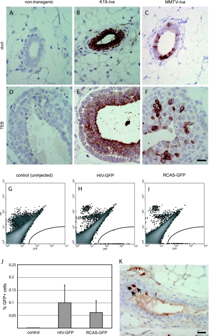
Figure 1.
Lentiviral infection in K19-tva transgenic mice. (A–F) Immunohistochemical staining for TVA in mammary gland sections from pubertal K19-tva transgenic mice. Staining results of both ducts and terminal end buds (TEBs) are shown. Genotypes are indicated at the top. Scale bar, 20 µm. (G–I) The left #2 to 4 mammary glands of six adult K19-tva transgenic mice were injected with HIV(ALSV)-GFP (104 IU/gland), and an equal amount of RCAS-GFP was injected into each of the contralateral glands. Infection efficiency was determined by FACS for GFP 5 days after injection. Shown are representative FACS plots of mammary cells from uninjected (G), HIV(ALSV)-GFP-injected (H), and RCAS-GFP-injected (I) K19-tva transgenic mice. No phycoerythrin (PE) was added to the cells; the PE channel (Y-axis) was used to segregate autofluorescing cells (high in both PE and GFP channels) from GFP+ cells (high GFP and no PE). (J) Quantitation of GFP+ cells 5 days after injection (n = 6 mice, mean ± SD). (K) Immunohistochemical staining for GFP in mammary gland sections from adult K19-tva transgenic mice harvested 4 days after injection of HIV(ALSV)-GFP (105 IU) into the ductal lumen. Arrows indicate positively stained cells. Scale bar, 20 µm.

