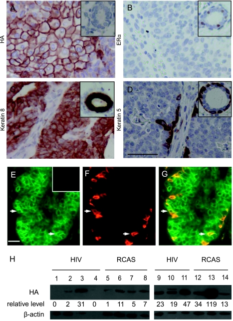
Figure 4.
Characterization of HIV(ALSV)-PyMT-induced mammary tumors. Paraffin sections from HIV(ALSV)-PyMT-induced tumors were analyzed by immunohistochemistry for HA-tagged PyMT (A), ERα (B), keratin 8 (C), and keratin 5 (D). In each case, the insert shows a representative staining of a normal mammary gland section. Scale bar, 25 µm; in all sections, n = 15 tumors. (E–G) HIV(ALSV)-PyMT-induced tumors contain myoepithelial cells of infected cell origin. Representative photomicrographs of coimmunofluorescent staining of paraffin sections from HIV(ALSV)-PyMT-induced mammary tumors for cells producing HA-tagged PyMT (E) and the myoepithelial marker keratin 14 (F). Arrows indicate myoepithelial cells positive for both keratin 14 and PyMT (G). Insert: negative HA staining in a tumor from an MMTV-Wnt-1 transgenic mouse. Scale bar, 25 µm; n = 5. (H) Western blot analysis of PyMT in tumors. Sample lysates from tumors induced by the indicated PyMT-expressing vectors were probed for the HA tag in PyMT (top panel), with β-actin serving as a loading control (bottom panel). Quantitation of relative HA signal intensity (corrected for total protein) is indicated below each lane (arbitrary units).

