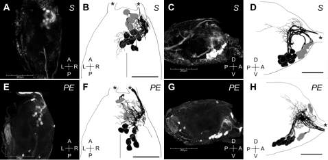
Figure 2. The mandibular motor neurons (MdMNs) stained by the retrograde tracing from the mandibular closer muscles of a soldier (A–D) and a pseudergate (E–H) in H. sjostedti.
The somata of MdMNs are located in the anterior region of the SOG and constitute the anterior cluster which contained 5 neurons (colored in grey), and the posterior cluster which contained 12 neurons (colored in black). Confocal images (A, E) and schematic images (B, F) in the dorsal view are shown. Anterior end of SOG facing up (A, B, E, F). Confocal images (C, G) and schematic images (D, H) in the lateral view are also shown. Anterior is right (C, D, G, H). Scale bars in A, C, E and G show 200 µm and those in B, D, F and H show 100 µm. Asterisks indicate mandibular nerves.

